Arm Muscles Diagram Posterior / : These septa divide the arm into its anterior and posterior compartments.. The muscles of the upper arm are used for the flexion and extension of the forearm at the elbow joint. Abducts the ulna when the arm is pronating (rotating forearm medially), helps triceps brachii extend forearm b. Want to learn more about it? 8 name the muscle of extensor compartment of arm and its nerve supply. Muscles in the posterior compartment of the forearm.
The arm muscles are located between the shoulder and elbow joint. For more anatomy content please follow us and visit our website: The superficial layer of the posterior compartment contains seven muscles that have a common origin of the supracondylar ridge and laterally epicondyle of the humerus (the. There will be plenty of other arm poses and practice activities to help improve. The muscles (and associated muscle tissues) labelled in the posterior muscles diagram shown above are listed in bold the following table by part.
Muscles Of The Forearm from antranik.org The muscles of this compartment are the triceps brachii and anconeus muscle and these are innervated by the radial nerve. Muscles of the posterior compartment of the forearm. Muscles of right upper arm. I will be breaking down each of these perspectives and showing how to draw the muscles, step by step. Posterior muscles of the arm and forearm. Learn more about their anatomy at kenhub! Posterior muscles of the body diagram (with images). Muscles of the arm and forearm.
These septa divide the arm into its anterior and posterior compartments.
Learn vocabulary, terms and more with flashcards, games and other study tools. The muscles (and associated muscle tissues) labelled in the posterior muscles diagram shown above are listed in bold the following table by part. I will be breaking down each of these perspectives and showing how to draw the muscles, step by step. This muscle diagram is interactive: Tutorials and quizzes on muscles that act on the arm/humerus (arm muscles: Muscles flexors in the arm all innervated the musculocutaneous nerve: The serratus posterior inferior muscle is thin but becomes more muscular at its lowest points. Their blood supply is from the profunda brachii. Mainly produce wrist and/or finger extension, and thumb abduction. The muscles of this compartment are the triceps brachii and anconeus muscle and these are innervated by the radial nerve. Muscles of the arm and forearm. Arm muscles diagrams diagram link human muscle anatomy arm muscle anatomy arm anatomy. The radial nerve is the largest branch of brachial plexus travels along with.
Human leg muscles diagram leg muscle chart gosutalentrankco. 7 draw labelled diagram showing branches of profunda brachi artery. For more anatomy content please follow us and visit our website: Posterior muscles of the arm and forearm. This muscle diagram is interactive:
Posterior Arm Muscles Diagram Quizlet from o.quizlet.com The muscles of the upper arm are split into anterior and posterior compartments. Muscles that cross the elbow (moving the forearm) (posterior view). The muscles of this compartment are the triceps brachii and anconeus muscle and these are innervated by the radial nerve. Draw labelled diagram showing branches of profunda brachi artery. Arm muscles with portions of arteries and nerves muscles of arm: 8 name the muscle of extensor compartment of arm and its nerve supply. Forearm muscles anatomy, posterior arm muscles, muscles of the arm and forearm, forearm anatomy, arm muscles diagram, deep muscles of forearm, muscles in lower arm. Mainly produce wrist and/or finger extension, and thumb abduction.
Related posts of shoulder muscles labelled diagram. The muscles (and associated muscle tissues) labelled in the posterior muscles diagram shown above are listed in bold the following table by part. Learn the muscles of the arm with free quizzes, diagrams and worksheets. File:muscle posterior labeled.png these pictures of this page are about:human muscle diagram posterior. Muscles of the arm and forearm. Muscles that cross the elbow (moving the forearm) (posterior view). Arm muscle diagram muscles of the rotator cuff human anatomy and physiology lab bsb 141. Muscles of right upper arm. Two intermuscular septa (medial and lateral) extend from it to attach to the humerus at the medial condylar ridge and lateral supracondylar ridge, respectively. Arm muscles diagrams diagram link human muscle anatomy arm muscle anatomy arm anatomy. Forearm muscles part posterior extensor compartment. Coracobrachialis brachialis biceps brachii coracobrachialis: 8 name the muscle of extensor compartment of arm and its nerve supply.
Flexion is accomplished through the brachialis, biceps brachii, and the brachioradialis. The posterior compartment of the arm is also known as the extensor compartment, as its main action is extension. 8 name the muscle of extensor compartment of arm and its nerve supply. File:muscle posterior labeled.png these pictures of this page are about:human muscle diagram posterior. Flexion of the forearm is achieved by a group of three on the posterior side of the arm the extensor muscles, such as the extensor carpi ulnaris and extensor digitorum, act as antagonists to.
Muscles Of The Neck And Torso Classic Human Anatomy In Motion The Artist S Guide To The Dynamics Of Figure Drawing from doctorlib.info It is supplied with blood by the lowest posterior intercostal artery, the subcostal right serratus anterior. Muscles flexors in the arm all innervated the musculocutaneous nerve: Click on the name of a muscle for a page about that muscle (works for most labels). Human leg muscles diagram leg muscle chart gosutalentrankco. From lateral epicondyle of the humerus d. Anatomynote.com found posterior muscles of the upper arm from plenty of anatomical pictures on the internet. There will be plenty of other arm poses and practice activities to help improve. Arm muscle diagram muscles of the rotator cuff human anatomy and physiology lab bsb 141.
It is supplied with blood by the lowest posterior intercostal artery, the subcostal right serratus anterior.
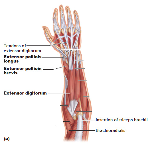
The muscles (and associated muscle tissues) labelled in the posterior muscles diagram shown above are listed in bold the following table by part. Lower left arm, posterior view, back of hand facing front. Abducts the ulna when the arm is pronating (rotating forearm medially), helps triceps brachii extend forearm b. Muscles that cross the elbow (moving the forearm) (posterior view). Muscles in the posterior compartment of the forearm. The muscles of the upper arm are used for the flexion and extension of the forearm at the elbow joint. The sacrum bone is almost always noticeable, no matter what the body type, because it is not covered with muscles or substantial fatty tissue. Muscles of the posterior compartment of the forearm. This class is focused on arm anatomy. Arm muscles diagrams diagram link human muscle anatomy arm muscle anatomy arm anatomy. Human leg muscles diagram leg muscle chart gosutalentrankco. Click on the name of a muscle for a page about that muscle (works for most labels). There will be plenty of other arm poses and practice activities to help improve.
Arm anatomy diagram for artists arm muscles diagram. Their blood supply is from the profunda brachii.


0 Komentar